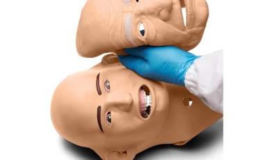
Dental Radiography Head Phantom
Features
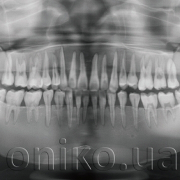
- Each hard tissue (enamel, dentin, cortical bone and cancellous bone) has particular HU number and X-ray absorption rate, each tooth has three-layer structure of enamel, dentin and pulp cavity
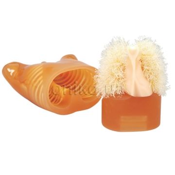
- Jaws and tongue are detachable to allow access to the oral cavity, pharyngeal cavity and maxillary sinus. Sensors, simulated lesions, or residue can be set in these cavities
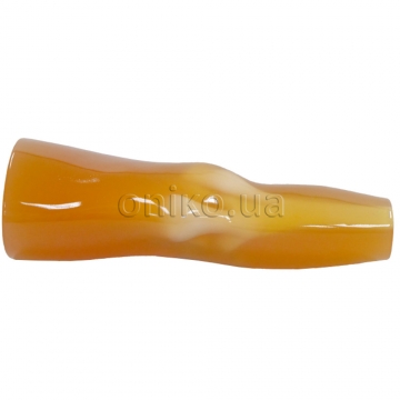
- Carotid arteries are prepared as lumens to accommodate simulated calcifications
Skills:
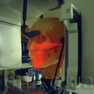
Tooth jaw X-ray training and quality assurance technique:
- Panoramic X-ray
- Intraoral X-ray
- Dental CT
- Cephalogram


